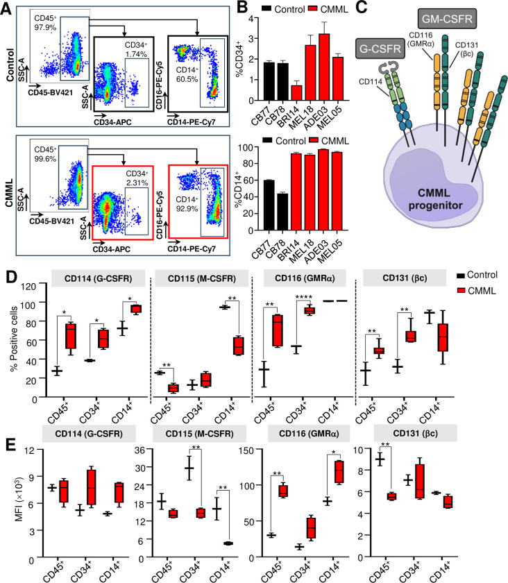
Fig 3. CMML have an increased percentage of CD116 and CD131 positive CD34+ stem and progenitor cells.
(A) Flow cytometry analysis of a representative CMML sample and healthy control stained for CD45, CD34, CD14 and CD16, and gating strategy used to define CD45+ mononuclear cells, CD34+ stem and progenitor cells and CD14+ monocytes. (B) Percentage of CD34+ progenitors and CD14+ monocytes in CMML samples (n = 4) vs. healthy control (n = 2). (C) Illustration of the cluster of differentiation (CD) markers where CD114 is a marker for G-CSFR, CD116 GMRα and CD131 βc. G-CSFR is homodimeric while GM-CSFR is heterodimeric receptor consisting of GMRα and βc. The expression of CD114, CD115, CD116 and CD131 in CMML samples (n = 4–6) vs. control (n = 2–3) in CD45+, CD34+ and CD14+ subpopulations, expressed as percentage of positively stained cells (D) and MFI (E) compared to control (cord blood or peripheral blood mononuclear cells from healthy donors). Bars represent mean ± standard deviation in (B). Box and whiskers graphs were plotted with min and max in (C) and (D). Unpaired Student’s t-test between CMML vs. healthy control used to determine statistical significance, where P<0.05 was statistically significant. *P<0.05, **P<0.01, ***P<0.001, ****P<0.0001. [MFI mean fluorescence intensity; G-CSFR granulocyte-colony stimulating factor receptor; GMRα granulocyte-macrophage colony stimulating factor receptor subunit α; βc beta common subunit].

