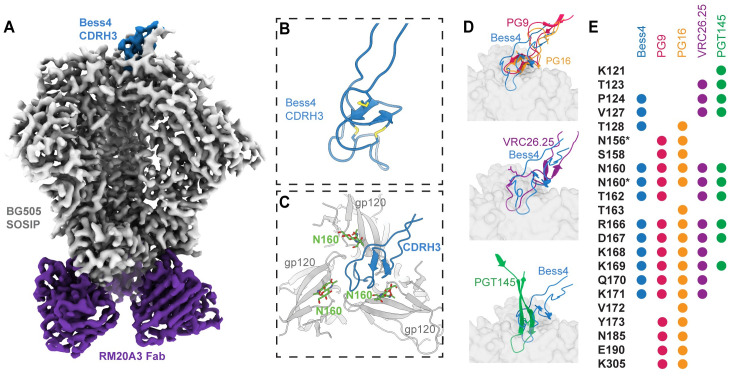
Fig 7. Cryo-EM structure of Bess4 in complex with BG505 SOSIP.
(A) 3.3 Å cryo-EM reconstruction of Bess4 Fab in complex with BG505 SOSIP and RM20A3 Fab (used to increase orientation sampling). (B) Atomic model of Bess4 CDRH3 backbone with 3 pairs of disulfide bonds highlighted. (C) View of CDRH3 interaction with BG505 SOSIP as viewed down the trimer apex 3-fold axis. The three gp120 protomer chains are labeled A, B and C. (D) Comparison of Bess4 CDRH3 with human bnAbs PG9, PG16, CAP256-VRC26.25 and PGT145. Viewing plane is perpendicular to the viral membrane, with trimer apex at the top of the Fig (E) Comparison of contact residues of BG505 SOSIP as defined by a buried surface area >10 Å2 between antibody CDRH3 and gp120. Residues with an asterisk denote contacts with N-linked glycan sugar(s).

