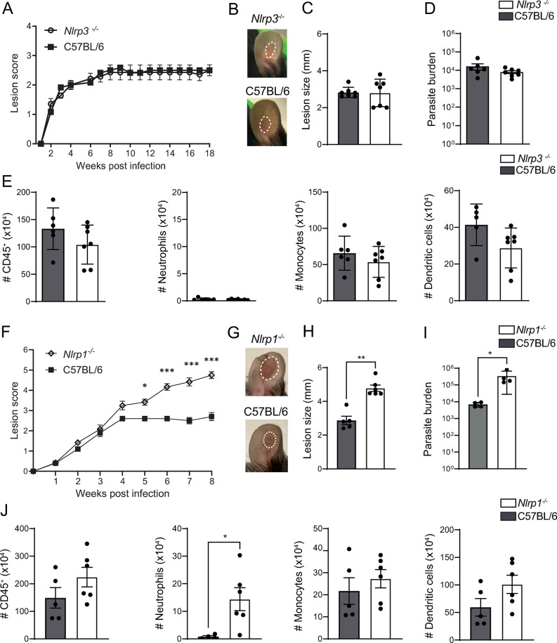
Fig 1. NLRP1, but not NLRP3, protects against lesion exacerbation during L.
mexicana infection. (A) Nlrp3-/- and C57BL/6 control mice were infected i.d. with 106 L. mexicana metacyclic promastigotes. Lesion development was measured weekly over the indicated time frame, and lesion score was determined including lesion size, inflammation, and pathology status. (B) Eighteen weeks p.i., representative lesion pictures are shown, (C) lesion size was measured and (D) parasite load at the site of infection was determined by limiting dilution assay (LDA). (E) The number of CD45+ cells and the relevant immune populations, including CD45+CD11b+Ly6G+ neutrophils, CD45+CD11b+Ly6C+ monocytes, and CD45+CD11c+ dendritic cells in the infected ear, was determined by flow cytometry. (F) Nlrp1-/- and C57BL/6 control mice were similarly infected i.d. with metacyclic L. mexicana promastigotes and lesion score was monitored over 8 weeks. (G) Representative pictures of Nlrp1-/- and C57BL/6 ear lesions at 8 weeks p.i. (H) Representative ear lesion size and (I) parasite load as determined by LDA. (J) The number of CD45+ cells, CD45+CD11b+Ly6G+ neutrophils, CD45+CD11b+Ly6C+ monocytes, and CD45+CD11c+ dendritic cells in the infected ears was analyzed 8 weeks p.i. by flow cytometry. Data are shown as mean ± SD and are representative of ≥3 experiments, n≥4/group. Statistical differences in lesion development were analyzed by 2-way ANOVA, and cell numbers using a Mann-Whitney U-test. *p <0.05; **p <0.01. ***p <0.001.

