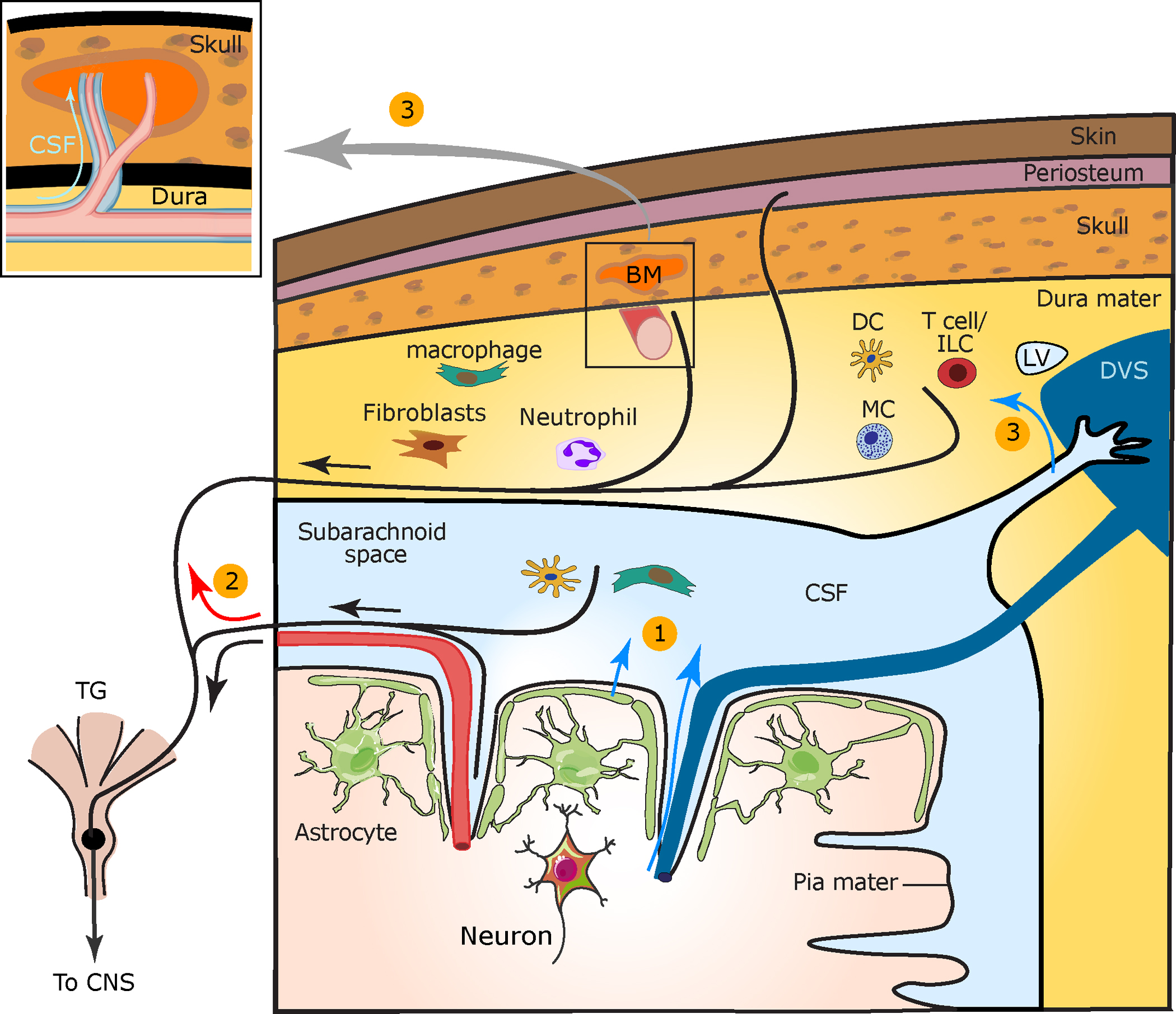
Figure 1.

Cortical to meningeal signaling underlying CSD-evoked meningeal nociception. CSD is a key event in migraine with aura. (①) In the wake of CSD, algesic mediators are released from cortical neurons and astrocytes and diffuse across the pial membrane or are transported via glymphatic flow into the CSF (blue arrows). These mediators may directly activate meningeal afferents surrounding pial vessels. Released cortical mediators may also activate subdural immune cells to release pronociceptive factors and excite pial afferents. (②) Antidromic axon reflex (red arrow) is posited to cause the release of neuropeptides from collateral dural afferent fibers that produce neurogenic inflammation, which involves vascular and immune responses with subsequent sensitization of dural afferents. (③) CSF algesic mediators are additionally transported into the dura mater near venous sinuses, lymphatic vessels, and perivascular spaces surrounding dural vessels (box) and may further sensitize dural afferents. CSF flow into calvarial bone marrow via channels may influence meningeal immune cell production and transport and locally trigger collateral afferents. Abbreviations: BM, bone marrow; CNS, central nervous system; CSD, cortical spreading depolarization; CSF, cerebrospinal fluid; DC, dendritic cell; DVS, dural venous sinus; LV, lymphatic vessel; MC, mast cell; TG, trigeminal ganglion. Figure adapted from Carneiro-Nascimento & Levy (2022) (CC BY 4.0).
