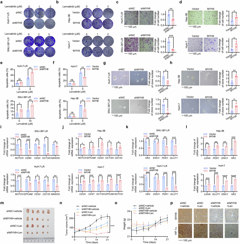
Fig. 4.
MYH9 promotes LR and cancer stemness in HCC. HCC LR cells with MYH9 knockdown (shMYH9) or shNC. a and WT HCC cells with MYH9 overexpression or vector (b) were cultured with the indicated lenvatinib concentrations. Following 14 days, crystal violet staining was used to determine the remaining cells. The influence of shNC and shMYH9 on HCC LR cell stemness was demonstrated through assessments of migration (c), flow cytometry (e), in vitro self-renewal (g) and the mRNA expression of stemness markers (i) and glycolysis driver genes (k). The influence of vector and MYH9 overexpression on WT HCC cell stemness was demonstrated through assessments of migration (d), flow cytometry (f), in vitro self-renewal (h) and the mRNA expression of stemness markers (j) and glycolysis driver genes (l). m HuH-7-LR cells with shMYH9 or shNC xenografted nude mice were treated with vehicle or Len (lenvatinib, 30 mg/kg) for 3 weeks after the tumor reached an average size of 50–100 mm3 (n = 5 per group). After 21 days, the mice were sacrificed. The isolated tumors were photographed. Tumor volume (n) and mouse weight of each group (o) were recorded every other week. p Immunohistochemistry was used to assess the expression of MYH9 and HIF-1α in the xenografts post-treatment. The data of cell functional assays and RT-qPCR analysis were presented as mean ± SD of three individual experiments, and the data of animal experiments were presented as mean ± SEM. The Student’s t-test was used for comparisons. *p < 0.05, **p < 0.01, ***p < 0.001, ****p < 0.0001. Scale bar: 100 μm., non-muscle myosin heavy chain 9; WT wild type, LR lenvatinib resistant, HIF-1α hypoxia-inducible factor-1α, HCC hepatocellular carcinoma, ns nonsignificant

