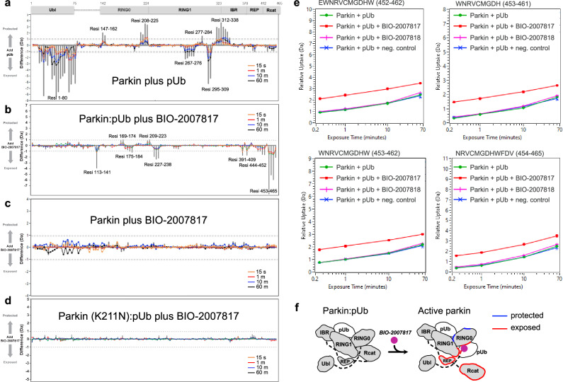
Fig. 3. HDX-MS analysis of conformational changes in parkin upon pUb and BIO-2007817 binding.
a Changes in solvent exposure of the parkin complex upon addition of pUb at four time points (15 s, 1 min, 10 min, 60 min) and the sum of the four time points (gray bars). The largest changes occur in the Ubl domain, which is released by pUb binding, and RING1, which is the site of pUb binding. b Changes in solvent exposure upon addition of BIO-2007817 in the presence of pUb. The largest changes occur in the Rcat domain. c, d Addition of BIO-2007817 does not change solvent exposure in the absence of pUb or in the parkin K211N mutant. e HDX-MS analysis of individual peptides from the parkin Rcat domain show solvent exposure in the presence of BIO-2007817 but not the inactive diastereomer BIO-2007818. f Mapping of increased (red) and decreased (blue) exchange upon BIO-2007817 addition to parkin:pUb (b).

