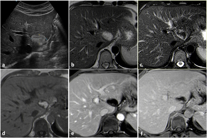
Fig. 8.
Angiomyolipoma. On B-mode US (a) a heterogeneous hyperechoic lesion is appreciable. The lesion appears markedly hyperintense on T2-weighted images (b) becoming prevalently hypointense after fat saturation (c) because of the macroscopic fatty content. On T1-weighted images (d) the lesion appears heterogeneously hyperintense; the peripheral components resemble a globular enhancement pattern during arterial (e) and portal venous (f) phases

