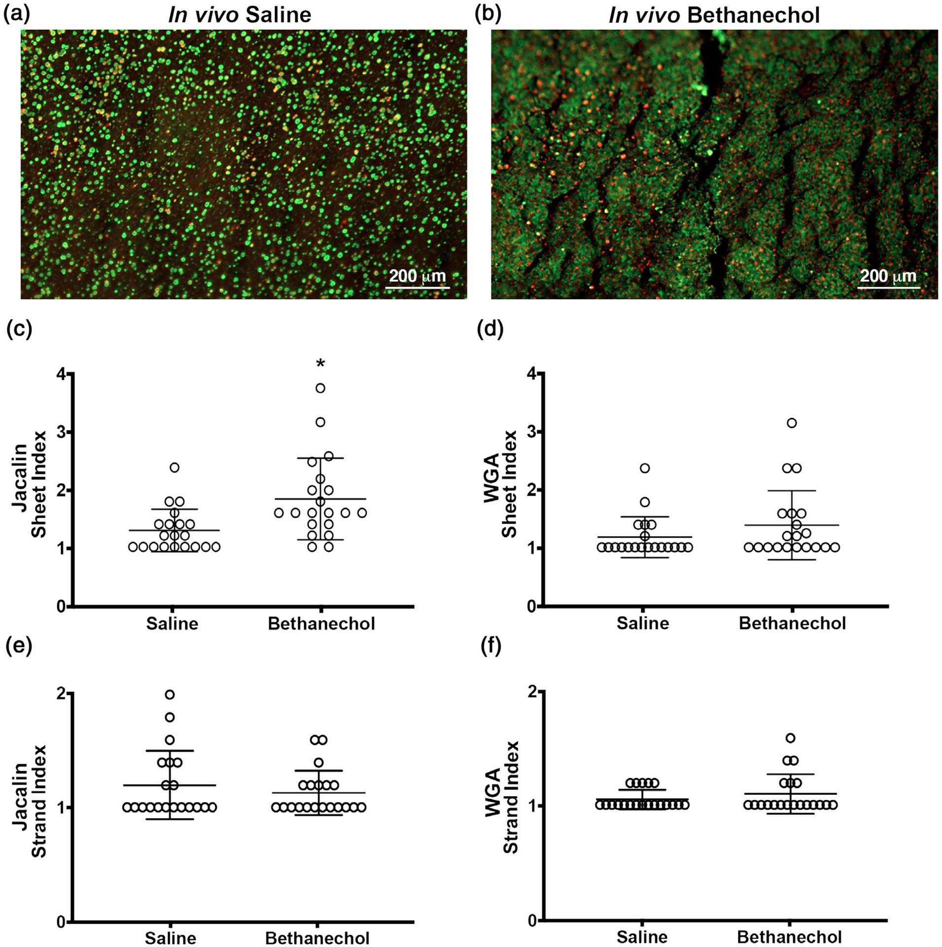
FIGURE 1.

Morphology of mucus in pig airways challenged with bethanechol and exposed 48 h later to the cholinergic agonist methacholine. (a,b) Representative en face image of an ex vivo trachea from a saline (a) or bethanechol (b) challenge after secondary exposure to the secretagogue methacholine. Discrete entities of mucus were observed and visualized by jacalin lectin (green) and WGA (red) staining. (c,d) Sheet index for jacalin-labelled mucus (c) and WGA-labelled mucus (d). (e,f) Strand index for jacalin-labelled mucus (e) and WGA-labelled mucus (f). n = 20 saline-challenged piglets (10 females and 10 males) and n = 20 bethanechol-challenged pigs (10 females and 10 males). Data points represent the mean score for each piglet calculated from five to seven images analysed (encompassing the anterior, lateral and posterior regions of the trachea). A greater score indicates greater incidence of the feature measured. Abbreviation: WGA, wheat germ agglutinin. *P < 0.05 compared with saline-challenged pigs. For panels e and f, data were assessed with a non-parametric Mann–Whitney U test. All data are shown as mean values ± SD
