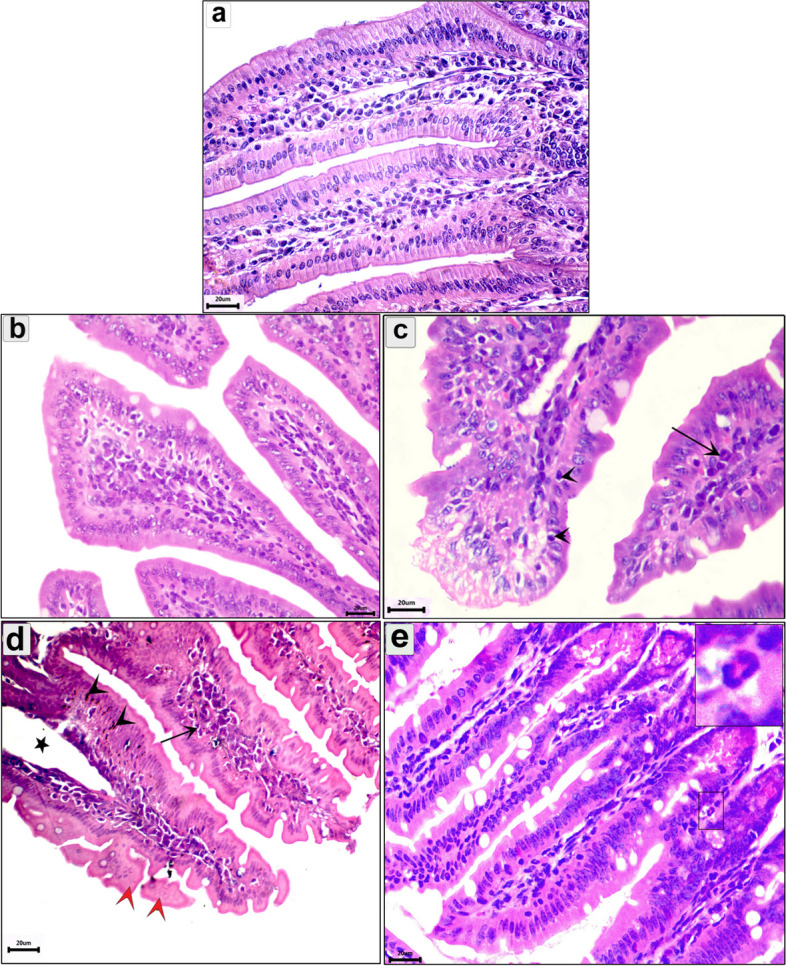
Fig. 3.

Photomicrograph of the intestine in the different groups in the enteral phase showing: a Normal structure of the intestinal mucosa; b uninfected pumpkin-treated intestine showing normal villi with healthy enterocytes; c Transverse section (T.s) in the intestine of the infected untreated group showing the parasitic enteritis characterized by shedding of the epithelium high inflammatory reaction in the core of the villi (arrow) and mononuclear cells between enterocytes (arrowhead); d T.s in the intestine of the pumpkin-treated group showing less normal intestinal mucosa, hyperplasia in some enterocytes (arrowhead), desquamation of the epithelium in tips of villi (red arrowhead), and mild inflammatory reaction in the core of villi (arrow); e T.s in the intestine of the albendazole-treated group showing normal intestinal villi with high number of goblet cells and eosinophils infiltration (small box). Stain H&E (400 ×)
