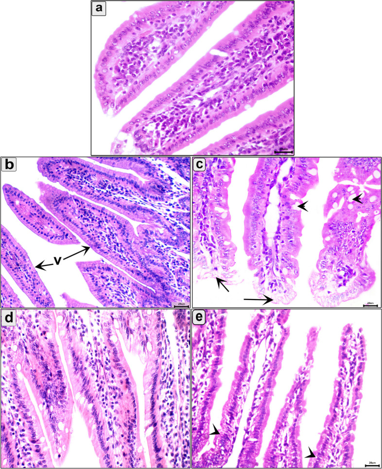
Fig. 4.

Photomicrograph of the intestine in the different groups in the parenteral phase showing: a Normal structure of the intestinal mucosa; b T.s in the intestine of uninfected pumpkin-treated showing normal villi (v) with healthy enterocytes; c T.s in the intestine of infected untreated group showing vacuolar degeneration in the enterocytes at the tips of villi (arroe) and increase the number of goblet cell (arrowhead); d T.s in the intestine of pumpkin-treated group showing normal architecture of intestinal mucosa; e T.s in the intestine of albendazole-treated group showing necrobiotic changes and vacuolar degeneration of enterocytes (arrowhead). Stain H&E (400 ×)
