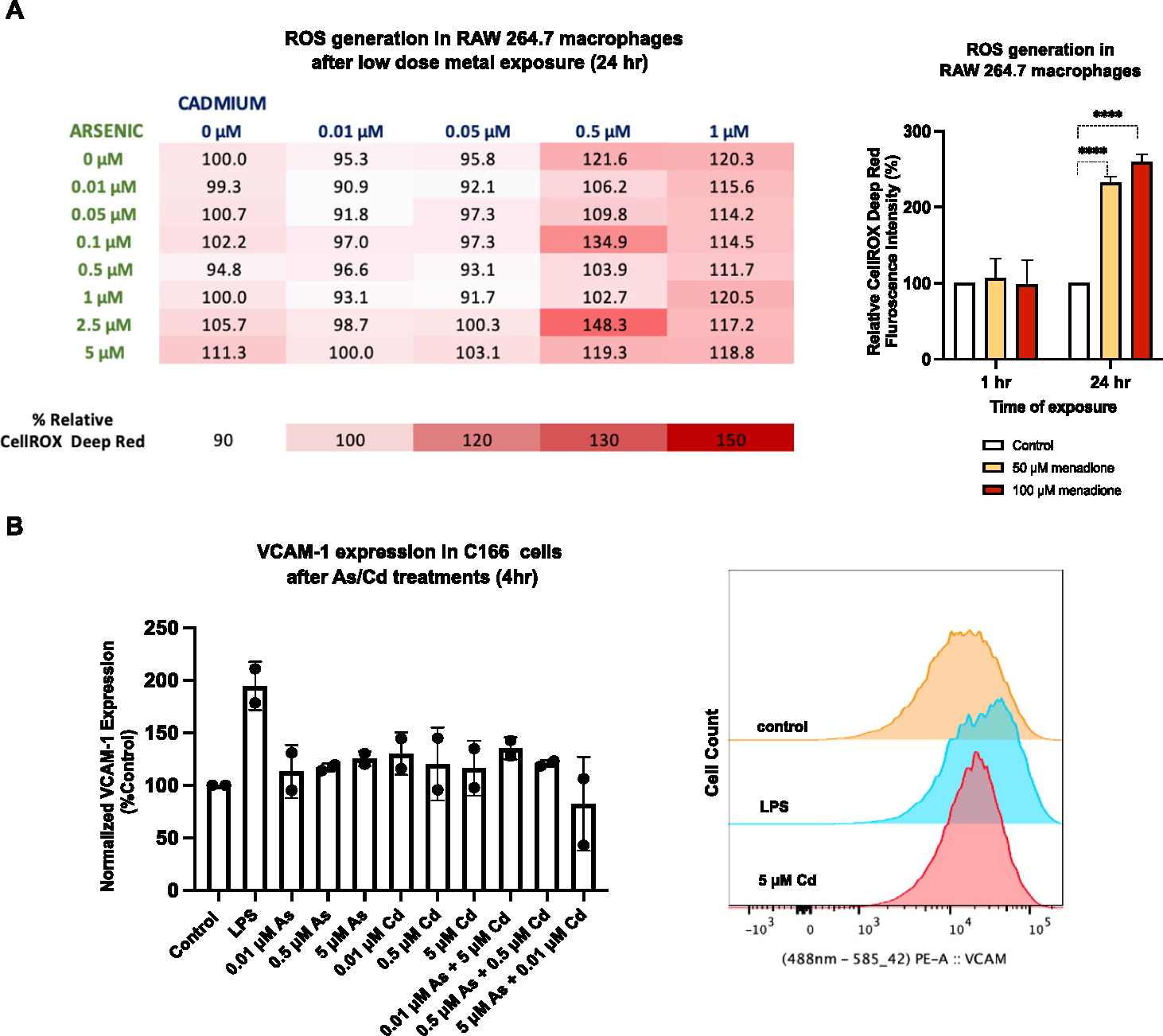
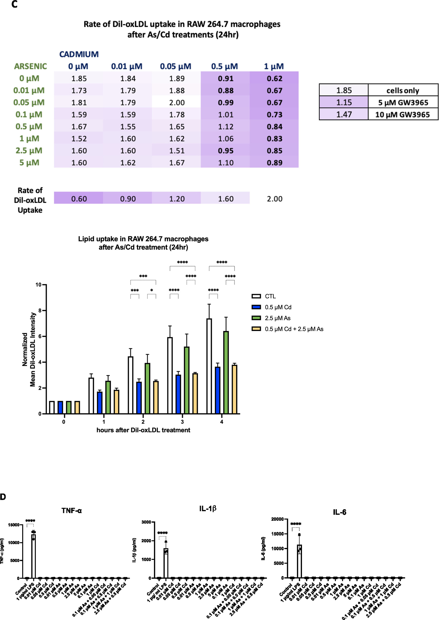
Fig. 2.


In vitro approaches to study atherosclerosis following exposure to low-dose arsenic and cadmium. (A) Intracellular ROS levels were measured and quantified as a change in CellROX Deep Red fluorescence signal by high content screening. Increased ROS levels are seen at higher concentrations of arsenic, cadmium and in combinations, although not statistically significant. Menadione was used as a positive control. Statistical significance is represented as follows: ****p < .0001. (B) Flow cytometry analysis of the expression of VCAM-1 on C166 cells under low dose-arsenic and cadmium treatment over 4 h (n = 2). No changes were observed in VCAM-1 expression after low-dose metal treatments. (C) Dil-oxLDL uptake was measured in RAW cells after 24-h metal exposure (n = 3). At higher concentrations of cadmium, the rate of Dil-oxLDL uptake (per hour) was significantly reduced (represented in bold) singularly and in combinations compared to cells only. A two-way ANOVA with a Tukey’s multiple comparison test was done. Statistical significance is represented as follows: *p < .05, **p < .01, ***p < .001 and ****p < .0001. (D) No change in TNF-α, IL-1β and IL-6 expression after exposure to arsenic and cadmium in RAW 264.7 cells. LPS was used as a positive control. (For interpretation of the references to colour in this figure legend, the reader is referred to the web version of this article.)
