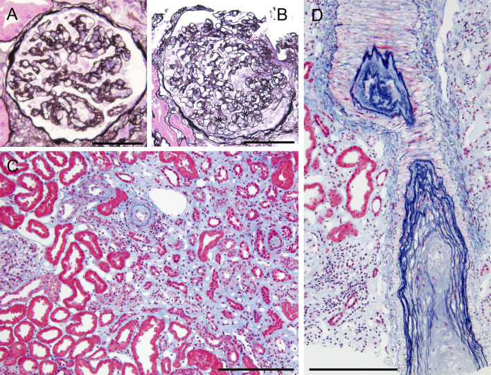
Figure 2.
Light micrographs of a kidney biopsy. A: A glomerulus shows capillary wrinkling, irregular double contours and collapse of capillary tufts. Periodic acid-methenamine silver staining. Original magnification ×400. Bar=100 μm. B: A glomerulus shows mesangiolysis (asterisks). Periodic acid-methenamine silver staining. Original magnification ×200. Bar=100 μm. C: There are mild tubular atrophy and interstitial fibrosis with edema, and distribution of the tubulointerstitial damage is zonal. Elastica-Masson staining. Original magnification ×200. Bar=100 μm. C and D: The small arteries and arterioles show marked intimal thickening with hyalinosis, indicating luminal narrowing or obstruction. Elastica-Masson staining. Original magnification ×200. Bar=100 μm.

