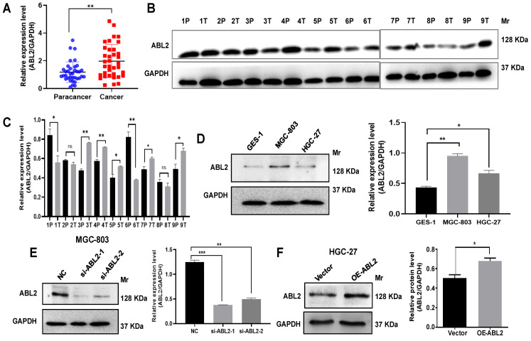
Figure 1.
ABL2 is overexpressed in GC tissues and cell lines. (A) ABL2 mRNA expression in 36 paired GC tissues and the corresponding paracancer tissues was detected by qRT-PCR. (B) Representative image shown detection of ABL2 expression in GC tissues (T) and corresponding paracancer tissues (P) from 9 GC patients by Western blotting assay. (C) Quantitative analysis of the relative ABL2 expression level in GC tissues. (D) Left, Representative image shown detection of ABL2 expression in GES-1 cells, MGC-803 cells and HGC-27 cells by Western blotting assay. Right, Quantitative analysis of the relative ABL2 expression level. (E) Left, Representative image shown detection of ABL2 expression in MGC-803 cells was knocked down by transfecting si-ABL2. Right, Quantitative analysis of the relative ABL2 expression level. (F) Left, Representative image shown detection of ABL2 expression by using Arg Lentiviral Activation Particles to overexpress ABL2 in HGC-27 cells. Right, Quantitative analysis of the relative ABL2 expression level. Data are presented as the mean ± SD of 3 independent experiments. *P<0.05, **P<0.01, ***P<0.001. Mr: Molecular size.

