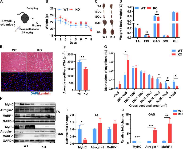
Fig. 3. SELENOW deletion aggravates DEX-induced muscle atrophy.
(A) Schematic showing the acute sarcopenia mice model established by the treatment of DEX in 8-week-old mice for 8 days. (B) Comparison of DEX treatment on body weight in WT and KO mice, n = 8. (C and D) Gross morphology mass analysis of TA, EDL, Gas, SOL, and QU in mice after DEX treatment, n = 8. (E) Representative H&E staining and Laminin staining of myofiber cross section of TA after DEX treatment; scale bars, 100 μm. (F and G) Related myofiber CSA and frequency of distribution for CSA of TA muscle after DEX treatment, n = 8. (H to J) Western blot analysis of protein levels of MyHC, Atrogin-1, and MuRF-1 in TA and Gas muscle. GAPDH was used as the loading control, n = 4. Data are means ± SEM; Student’s t test, *P ≤ 0.05, **P ≤ 0.01, ***P ≤ 0.001.

