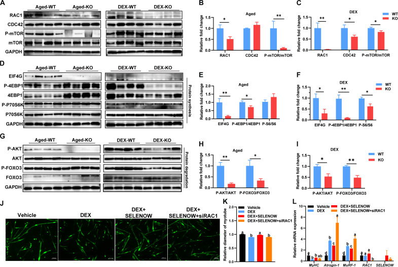
Fig. 6. SELENOW deletion effects on proteostasis via the RAC1-mTOR cascade.
(A to C) Western blot analysis of protein levels of RAC1, AT1, CDC42, mTOR, and P-mTOR (S2248) in GAS muscle of the sarcopenia mice model, GAPDH was used as the loading control, n = 4. (D to F) Western blot analysis for the protein synthesis pathways [EIF4G, 4EBP1, P-4EBP1(T45), P70S6K, and P-P70S6K (S434)] in GAS muscle of the sarcopenia mice model, GAPDH was used as loading control, n = 4. (G to I) Western blot analysis for the protein degradation pathways [AKT, P-AKT (S473), FOXO3, and P-FOXO3 (S253)] in GAS muscle of the sarcopenia mice model; GAPDH was used as the loading control, n = 4. (J) Immunofluorescent staining of MyHC in primary myotubes. Before DEX treatment, the primary myotubes were infected with a control vehicle, a SELENOW vehicle, or a combination of SELENOW vehicle and siRAC1 mimic; scale bar, 100 μm. (K) Quantification of these primary myotube diameters was performed, with at least 500 myotubes measured in each group. (L) Expression of MyHC, Atrogin-1, MuRF-1, RAC1, and SELENOW in the mRNA level of these primary myotubes; GAPDH was used as the loading control, n = 5 to 6. Data are means ± SEM; Student’s t test, *P ≤ 0.05, **P ≤ 0.01; labeled means without a common letter differ; P < 0.05.

