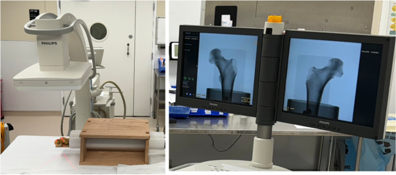
Figure 1.
Experimental Setup: The cadaveric bone specimen was positioned on a radiographic table in a manufactured rotation device made of a radiopaque plastic pipe and a box. A C-arm fluoroscopy machine (Phillips Zendition 70, Eindhoven, NL) was centered 12 inches above the femoral head for imaging of the Lesser Trochanter (LT) profile. Observers rotated the pipe at the distal end to adjust rotation angle of the bone until they were satisfied that they had matched the reference image.

