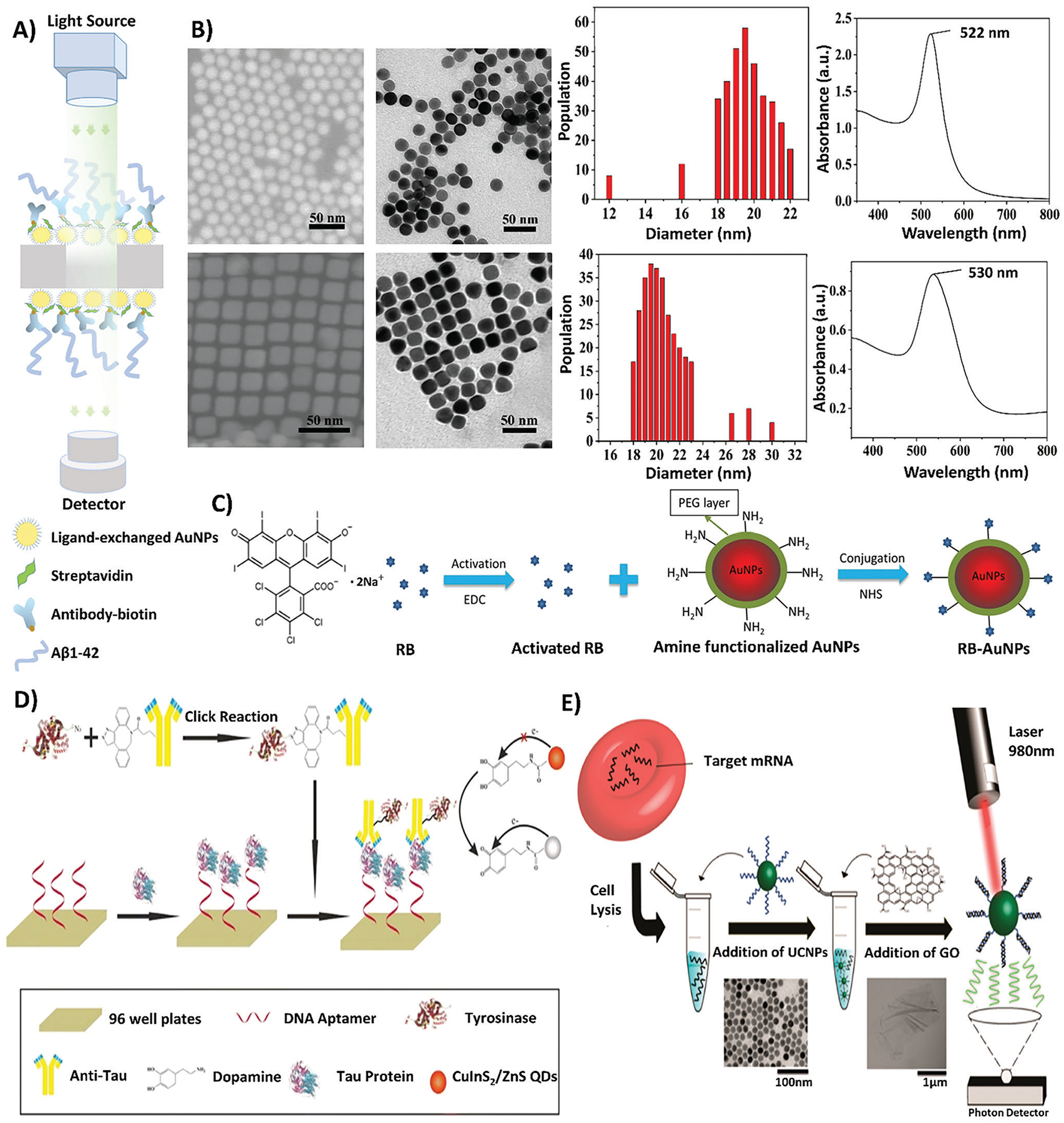
Figure 4.

A schematic of various optical biosensing measurement and fabrication techniques. A) A schematic depicting the utilization of LSPR quantification techniques to measure the optical properties of AuNPs in the biosensing platform. Reproduced with permission.[65] Copyright 2020, Elsevier. B) Depicts the characterization of AuNSs and AuNCs in terms of shape and absorbance. Reproduced with permission.[69] Copyright 2019, American Chemical Society. C) A schematic showing the conjugation process of the F-SERS probe, where RB is activated using EDC and combined with PEGylated AuNPs and NHS for conjugation, forming RB-AuNP complexes. Reproduced with permission.[75a,b] Copyright 2019, Springer Nature. D) A schematic outlining the application of CuInS2/ZnS QDs within a sandwich immunoassay for the fabrication of an optical biosensor. The process of creating the sandwich immunoassay involves the immobilization of DNA aptamers to a 96-well plate substrate, followed by the introduction of tau protein, and ending in the binding of anti-tau antibody conjugated to tyrosinase. Through dopamine-functionalization induced by tyrosinase enzymatic activity, the quenching effects of CuInS2/ZnS QDs are induced. Reproduced with permission.[83] Copyright 2019, Springer. E) A depiction of UCNPs in the detection of AD miRNAs, in which target miRNAs are extracted from cells through cell lysis and combined with UCNPs. With the addition of graphene oxide and the utilization of a 980 nm laser, the excitability of UCNPs with graphene oxide can be detected by a photon detector. Reproduced with permission.[84] Copyright 2017, American Chemical Society. The labels on all original figures have been modified to improve readability.
