Fig. 1.
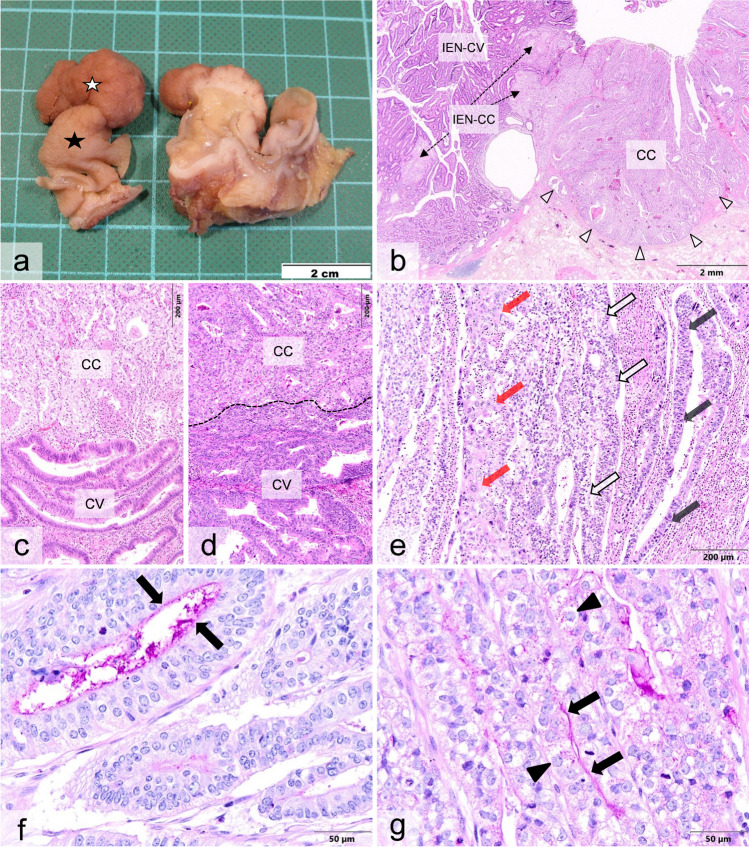
Macroscopical, histo-, and cytomorphological findings in colorectal adenocarcinoma in conventional (CV) and clear cell (CC) components in case 3 (a, b, c, f, g), case 1 (d), and case 2 (e). a Macroscopic finding in the formalin-fixed specimen. The polypoid tumor exhibits a homogenous brownish color (white star) on the external surface and a light brown to white color on the cut surface. The unaffected mucosa has a light brown color (black star). b Conventional tubulovillous adenoma (IEN-CV) as precursor lesion, which has progressed into a well-differentiated adenocarcinoma (not shown). The adenoma contains foci of tumor glands with clear cell changes (IEN-CC). CC component infiltrates the submucosa. Arrowheads point to invasion front. c CV and CC components, which are both low-grade differentiated appear sharply demarcated from each other with pushing margins. d Merging of CV and CC components, which are both high-grade differentiated. Dashed line denotes the blurred boundary of both components. e Tumor area with different patterns consisting of conventional (light gray arrows), clear cell (white arrows), and undifferentiated hepatoid types (red arrows). f, g PAS stains decorating the luminal cell membrane in CV (f) and CC component (g) marked by arrows. The tumor cells of the CC component are characterized by a lightened cytoplasm and contain depolarized enlarged nuclei with distinct nucleoli. Some clear cells contain fine PAS-positive granular deposits (arrowheads)

