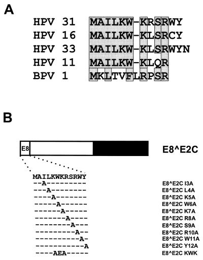
FIG. 4.
Alanine-scanning mutagenesis of the conserved E8 domain. (A) Alignment of E8 domains from HPV11, -16, -31, and -33 and BPV1. Identical or similar residues are boxed. (B) Structure and sequences of mutated HPV31 E8̂E2C proteins. The different domains of the E8̂E2C protein are shown at the top. The amino acid sequence of E8 residues 1 to 12 is depicted below, and the mutated residues are indicated.

