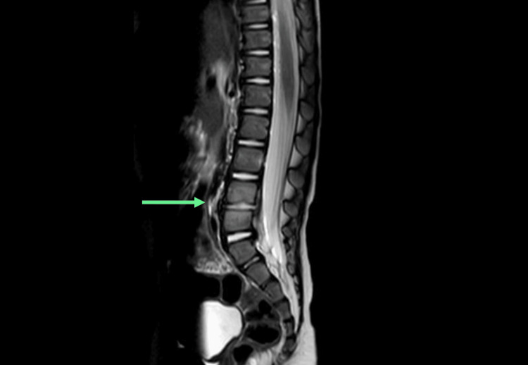
Figure 4. Sagittal view of an MRI of the lumbosacral spine. An arrow points to the disc between the L4 and L5 vertebrae. Fluid signal within the disc with minor loss of disc height and early end plate erosion of L4 and L5 vertebrae. Infective material/pus remains confined within the disc annulus, with no epidural hematoma or canal narrowing seen.
MRI: magnetic resonance imaging

