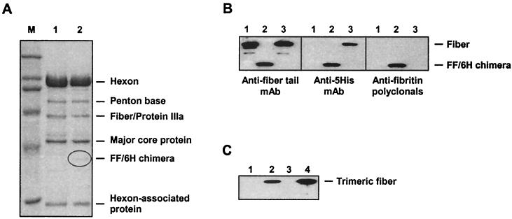
FIG. 2.
Analysis of Ad5LucFF/6H capsid composition. (A) SDS-PAGE of CsCl-purified Ad5LucFF/6H virions. Samples containing 4 × 10¹⁰ particles of either the wild-type Ad5 (lane 1) or Ad5LucFF/6H (lane 2) were boiled in Laemmli sample buffer and resolved on an SDS–10% PAGE gel. Of note, the resolution of this minigel is not sufficient for separation of the fiber and protein IIIa, which comigrate in lane 1. (B) Western blot analysis of FF/6H chimeras incorporated into Ad5LucFF/6H virions. Proteins of Ad5LucFF/6H virions denatured by being boiled in sample buffer (lanes 2) were separated on an SDS–10% PAGE gel and then probed with the anti-Ad fiber tail MAb 4D2, the anti-five-His MAb Penta-His, and anti-fibritin mouse polyclonal antibodies. Wild-type Ad5 (lanes 1) and Ad5LucFc6H, a virus containing wild-type fibers with carboxy-terminal six-His tags (lanes 3), were used as controls. (C) Western blot of Ad5Luc1 and Ad5LucFF/6H virions with anti-fiber knob antibody. Virions denatured by incubation in a sample buffer containing SDS were resolved on an SDS–10% PAGE gel and incubated with the anti-Ad5 fiber knob MAb 1D6.14 (9), which recognize the knob trimer. Lanes 1 and 3, Ad5LucFF/6H (3 × 10⁹ and 6 × 10⁹ virus particles per lane, respectively); lanes 2 and 4, Ad5Luc1 (same amounts of the virus as in lanes 1 and 3). Detection of viral proteins was done with the ECL Plus kit from Amersham Pharmacia Biotech (Piscataway, N.J.).

