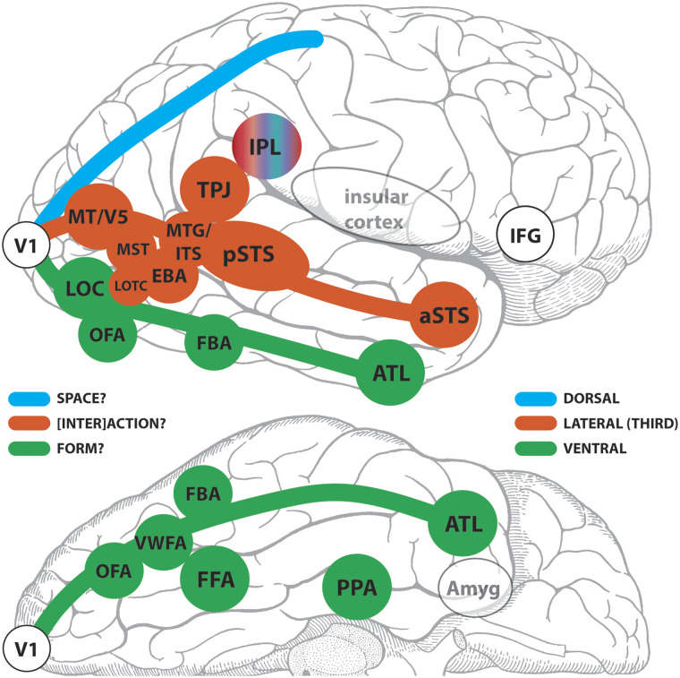
Figure 11. .
Putative members of the ventral and third/lateral pathways in the human brain? A schematic of a lateral (top) and partial inferior (bottom) view of a human cerebral hemisphere segregating some selected known functional brain areas into third/lateral or ventral pathways. The dorsal pathway appears as an outline and is not considered further here. The green, red-brick, and blue colors identify the three pathways for which primary visual cortex (V1) is the departure point. The inferior parietal lobule (IPL) is presented as an outlier and hence does not fit the color scheme. V1 = primary visual cortex; VWFA = visual word form area; MT/V5 = motion-sensitive fifth visual area; MST = human homolog of macaque medial superior temporal area with high-level motion sensitivity; MTG/ITS middle temporal gyral and inferior temporal sulcal cortex (sensitive to motion of animals and tools); TPJ = temporoparietal junction; Amyg = amygdala.

