
Figure 3.
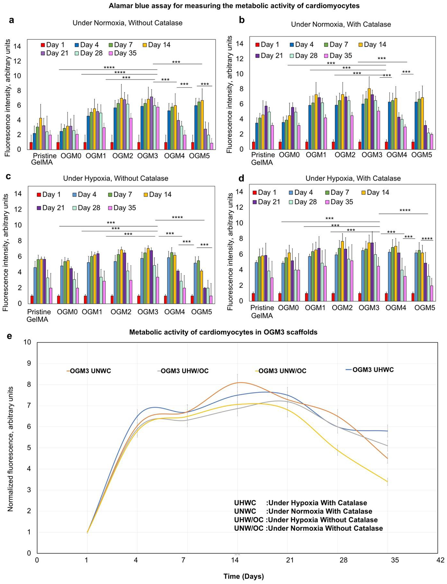
Cellular metabolic activity of encapsulated H9c2 cardiomyocytes evaluated using Alamar Blue assay a. Under normoxia without catalase, b. Under normoxia with catalase, c. Under hypoxia without catalase, and d. Under hypoxia with catalase; e. The OGM3 scaffolds displayed the highest metabolic activity over 35 days across all culture conditions. The comparative analysis of the metabolic activity within the OGM3 scaffolds was performed under all culture conditions. The OGM4 and OGM5 scaffolds caused a decline in the cellular metabolic activity despite their high oxygen content, indicating excessive oxygen concentrations decrease the cellular metabolic activity.