Figure 1.
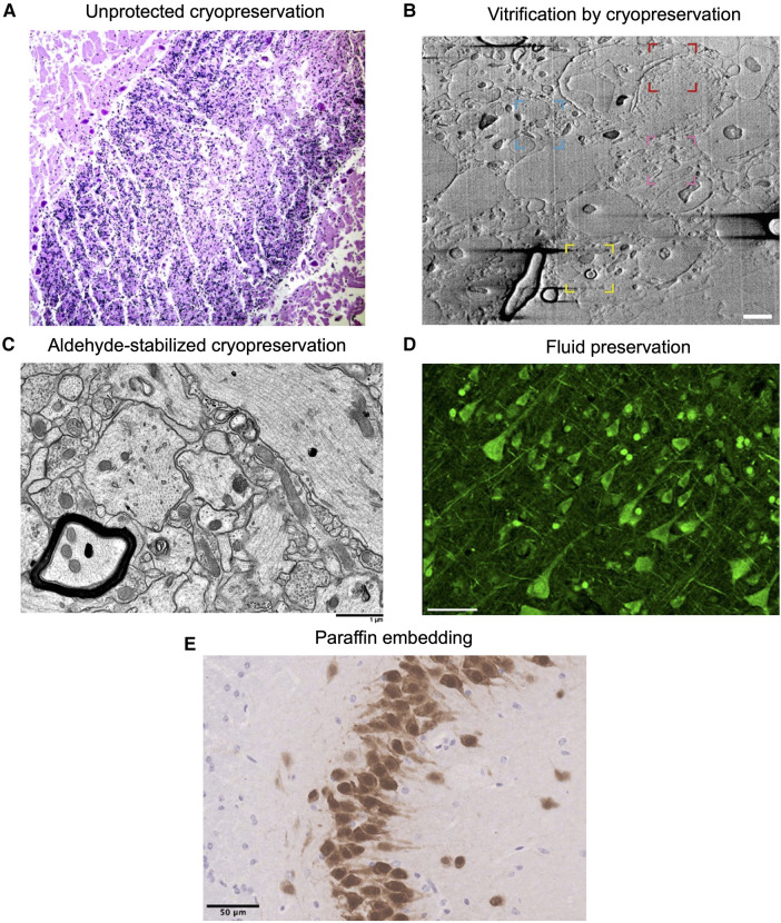
Example images showing morphology preservation in brain tissue preserved with different methods. (A) Image from (56). Crisscross linear clefts due to ice artifact in bovine cerebellar tissue, because of freezing and thawing without cryoprotectant. (B) Image from (57). Focused ion beam scanning electron microscopy (FIB-SEM) of a 200 µm brain tissue section cryoprotected in 20% bovine serum albumin and vitrified using high-pressure freezing, demonstrating preservation of myelin (yellow region), potential nuclear pore complexes (red), mitochondria (blue), and a synapse (pink). Scale bar: 1 µm. (C) Image from (53). Electron microscopy of rabbit brain fixed with 3% glutaraldehyde, cryoprotected with 65% ethylene glycol, vitrified, rewarmed, and cryoprotectant removed, demonstrating well-preserved structures. Scale bar: 1 µm. (D) Image from (58). Formalin-fixed human cortical tissue stained for pan-axonal neurofilaments with SMI312 after storage in fixative for 25 years, demonstrating intact neurons. Scale bar: 50 µm. (E) Image from (59). Perfusion-fixed mouse brain tissue that was dissected, paraffin embedded, and stained with NeuN, demonstrating expected neuronal morphology. Scale bar: 50 µm. All images reproduced under a Creative Commons license, available here: https://creativecommons.org/licenses/by/4.0/.

