
Figure 5.
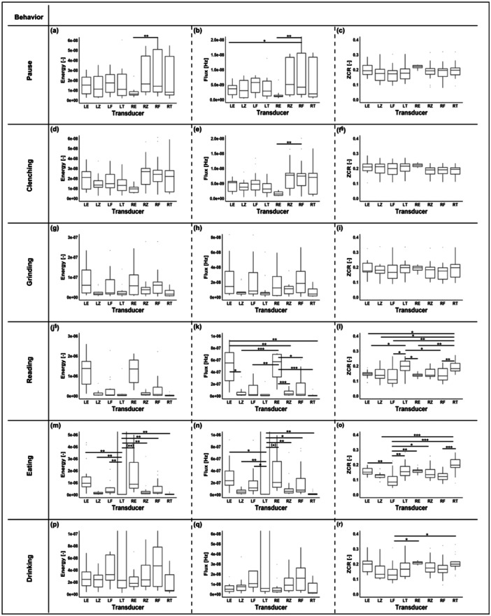
Energy, flux, and ZCR of all participants for each transducer and each behavior: clenching (a–c), grinding (d–f), reading (g–i), eating (j–l), drinking (m–o), and pause (p–r). The transducers are depicted on the horizontal axis as follows: left ear (LE), left zygomatic (LZ), left frontal (LF), left temporal (LT), right ear (RE), right zygomatic (RZ), right frontal (RF), and right temporal (RT). The marks inside the boxes represent the median, and the bottom and top of the boxes represent the 25th (q1) and the 75th (q3) percentiles, respectively. Significant differences between the different transducer placements are indicated with * for p < 0.05, ** for p < 0.01, and *** for p < 0.001. A full‐range illustration of figures (j), (k), (m), and (n) can be found in Supporting Information S1: Figure 1. §: Showed a significant difference as listed in Table 1 but was not included in the pairwise tests. The p values for the pairwise tests are listed in Supporting Information S1: Table 2. Note: (**) and (*) were added to two subfigures (m and n); they refer to instances where an additional nonparametric test was performed and the result was opposite to that of the ANOVA.