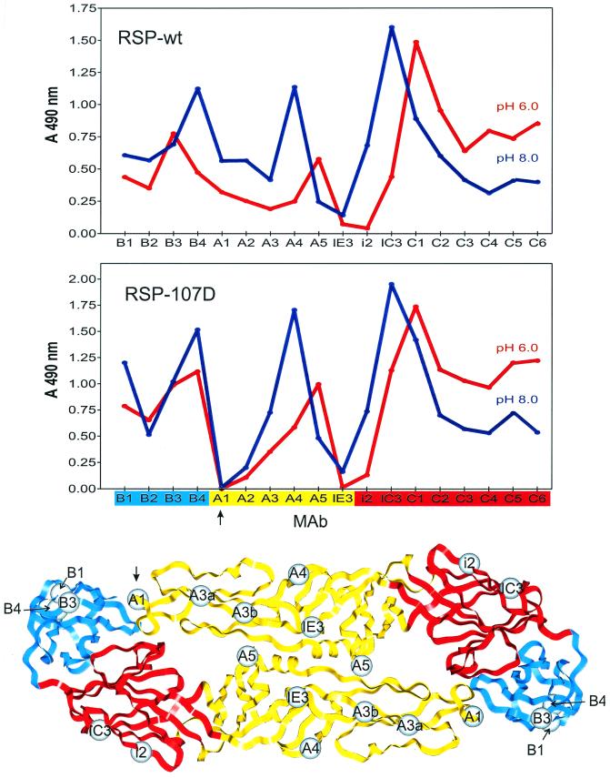
FIG. 4.
Antibody binding profiles before and after low-pH treatment. The binding activities of 18 E-protein-specific MAbs with low-pH-treated and untreated RSP-wt (upper panel) and RSP-107D (middle panel) were compared by four-layer ELISA. The blue patterns were obtained with untreated samples, and the red patterns were obtained with samples that had been preincubated at pH 6.0 and back-neutralized. The colors on the x axis of the middle panel represent the structural domains (depicted in the lower panel) to which the antibodies bind (red, domain I; yellow, domain II; blue, domain III). The spheres show the positions of mutations defining individual MAb binding sites that have been mapped by selection of neutralization escape variants of TBE virus (29) or mutations in RSPs that abolish binding of an individual MAb (A1 [this study] or B3 [unpublished data]). The epitopes for the nonneutralizing MAbs B2 and C1 to C6 have not been precisely mapped, but the antigenic domains to which they belong were established previously (29). Note that binding to MAb A1 (arrows) is abolished by the Leu 107-to-Asp mutation.

