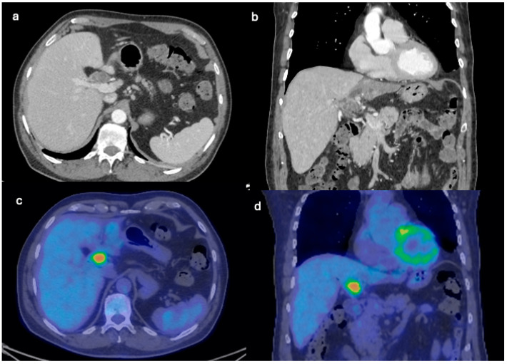
Figure 5.
CT axial and coronal planes on parenchymal window showing a multicystic lobulated mass involving the hepatic hilum. After injection of iodinated contrast, the mass appears hypo-vascular, with enhancing margins and internal septations (a,b). PET axial and coronal images show increased uptake of 18[F]-fluorodeoxyglucose in the multicystic lobulated mass involving the hepatic hilum suspicious for recurrences of IPBN (c,d).

