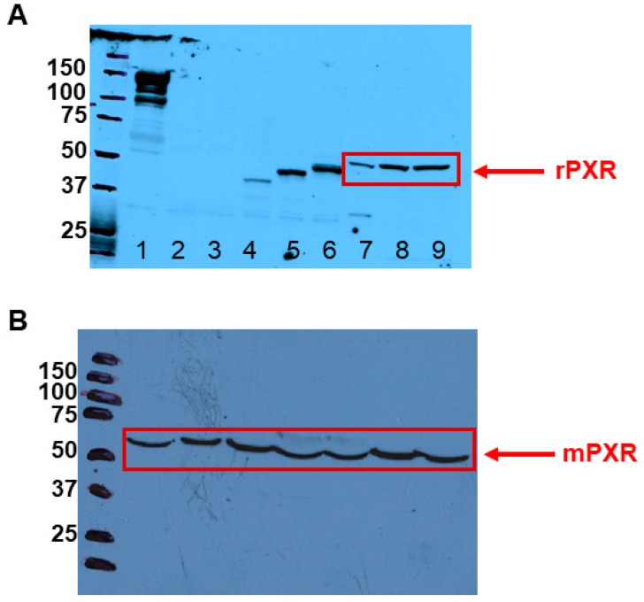
Figure 1.
PXR protein expression in rodent LCs. (A). Protein expression of rPXR in rat primary LCs was determined by Western blotting analysis (n = 2). Image shown is from a representative experiment. Lane 1, dog liver; Lane 2, COS-7 cells transfected with pcDNA; Lane 3, COS-7 cells transfected with FLAG-pcDNA; Lane 4, COS-7 cells transfected with human PXR (hPXR); Lane 5, COS-7 cells transfected with FLAG-hPXR; Lane 6, COS-7 cells transfected with 3XFLAG-hPXR; and Lanes 7 to 9, rat primary LCs. Marker molecular weights represent KDa. (B). Protein expression of mPXR in mouse MA-10 cells was determined by Western blotting analysis (n = 2). Image shown is from a representative experiment. All Lanes (1 to 7), MA-10 cells. Marker molecular weights represent KDa.

