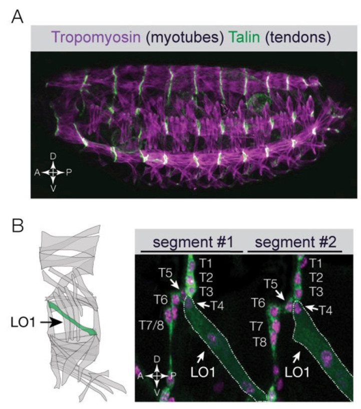
Figure 1.
Myotube guidance ensures muscles are targeted to the correct tendon. (A) Confocal micrograph of a Drosophila embryo near the end of embryogenesis (Stage 16) labeled for Tropomyosin (myotubes, violet) and Talin (tendon cells, green). Notice the musculoskeletal pattern is precisely repeated in each hemisegment along the anterior–posterior axis. (B) Live Stage 16 embryo that expressed nuclear RFP and membrane-bound GFP in all tendon cells and in a subset of myotubes. Eight tendon cells (T1-T8) and one LO1 myotube were labeled (dotted white line) in two adjacent hemisegments. The LO1 myotube attached to tendon T4 in both segments despite close proximity to seven other tendon cells. Diagram shows the 30 myotubes per embryonic segment. The LO1 myotube (Longitudinal Oblique 1) is shown in green.

