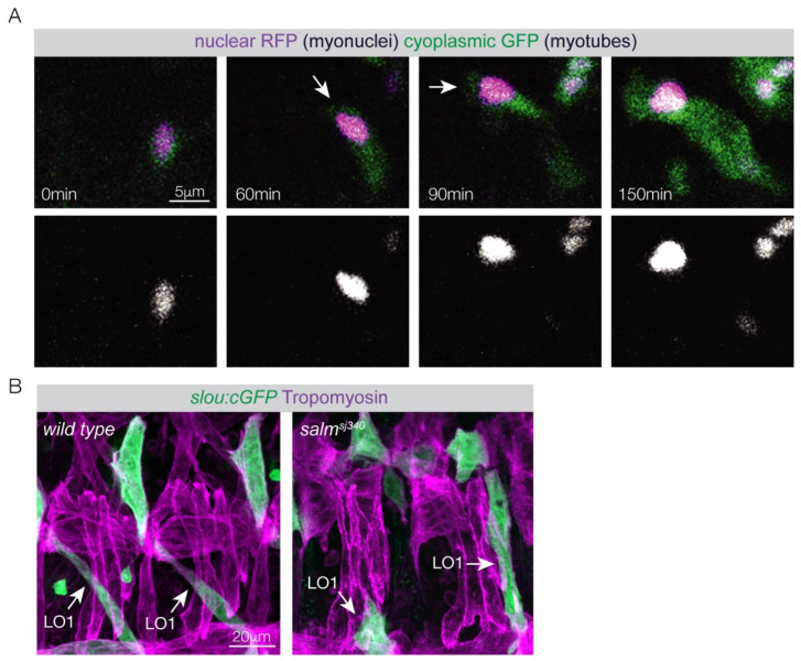
Figure 2.
Myotube guidance. (A) Live imaging of an LO1 myotube that expressed cytoplasmic GFP and nuclear RFP. Arrows highlight the dorsal myotube leading edge. At 60 min the dorsal myotube leading edge reached a choice point and navigated to a muscle attachment site on the anterior of the segment. A single myonucleus followed the leading edge throughout elongation (bottom row). Notice that myoblast fusion incorporates a second nucleus at 150 min. (B) Confocal images of Stage 16 embryos expressing the identity gene reporter slouch:GFP labeled for cytoplasmic GFP (green) and Tropomyosin (violet). The pattern of slouch:GFP myotubes was disrupted in salm mutant embryos. LO1 muscles often attached to the incorrect tendons.

