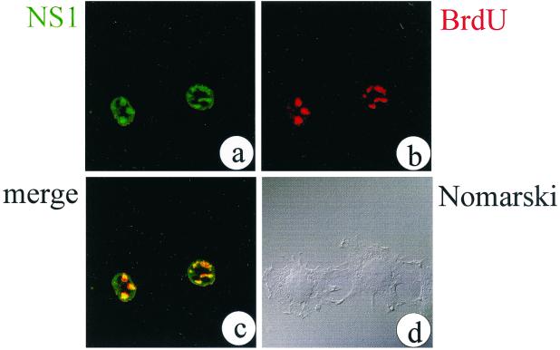
FIG. 1.
MVM DNA replication colocalizes with NS1 in APAR bodies in the nuclei of infected A9 cells. Representative confocal optical sections through the nuclei of infected cells are shown. NS1 was localized with the SP8 polyclonal antiserum and a fluorescein isothiocyanate (FITC)-conjugated secondary antibody (a). Replication was monitored by incorporation of BrdU and indirect immunofluorescence using a BrdU-specific antibody and a tetramethyl rhodamine isothiocyanate (TRITC)-conjugated secondary antibody (b). In a merged image, colocalized structures from panels a and b appear yellow (c). By phase-contrast microscopy (Nomarski), the cells show no obvious sign of NS1-induced cytotoxicity at the time of fixation (15 h postinfection) (d).

