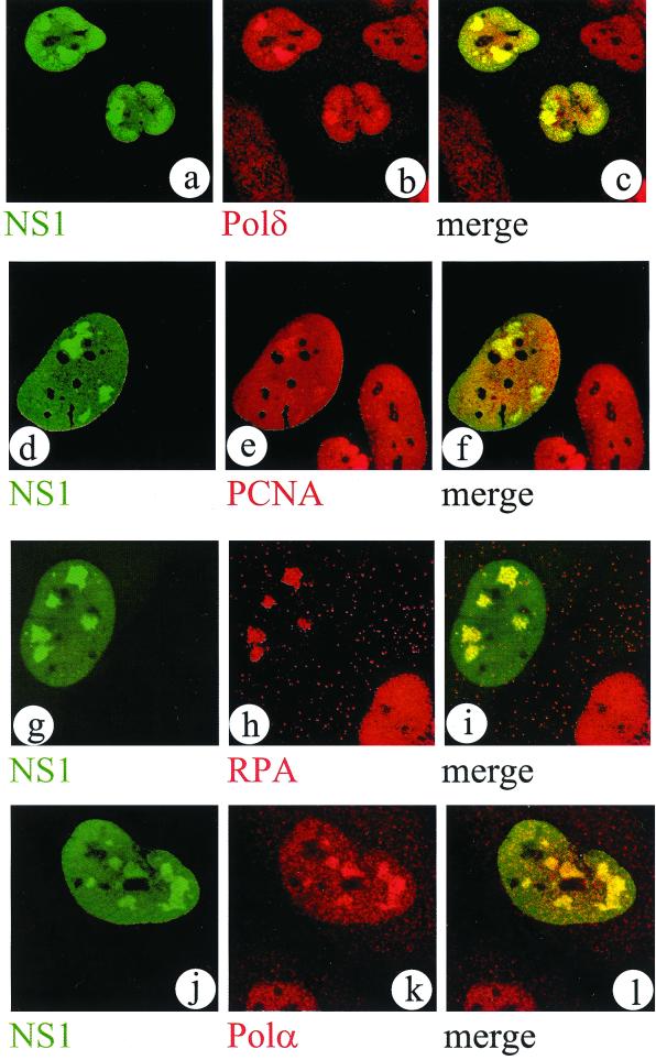
FIG. 3.
Cellular replication factors DNA polymerases δ and α, PCNA, and RPA accumulate in APAR bodies. Representative confocal images of nuclei from double-labeled MVM-infected NBE cells are shown. NS1 was detected with FITC using the SP8 antiserum (a, d, g, and j). Replication factors were detected with TRITC in the same confocal plane as shown in the left column, using the respective antibodies against DNA polymerase δ (Transduction Laboratories) (b), PCNA (PC10; Upstate Biotechnology) (e), RPA (Ab-1; Oncogene) (h), and DNA polymerase α (SJK132-20 [26]) (k), respectively. A merged image of both channels from the left and center columns provides evidence for colocalization of these factors in APAR bodies (c, f, i, and l).

