
Figure 2.
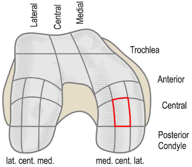
Femoral articular cartilage surface zones. The investigation site, medial femoral condyle central-central weight-bearing zone, is marked red. The weight-bearing region of the medial femoral condyle was visualized by flexing the knee to 45° and applying valgus pressure during arthroscopy.