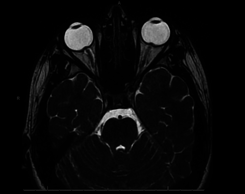
FIG. 1.
Axial T2 three-dimensional fast spin echo orbit sequence demonstrating bilateral posterior scleral flattening, prominent optic papillae at the site of optic nerve insertion, tortuosity of the intraorbital optic nerves, and prominence of the optic nerve sheath complex suggestive of papilledema and increased ICP.

