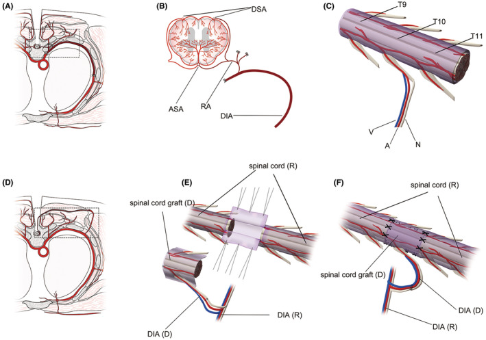
FIGURE 1.

Key surgical procedures and anatomy of vASCT. Cross‐sectional illustration of the T10 level of the donor before isolating the graft (A). Cross‐sectional view and 3D imaging depicting the spinal cord graft from the donor (B, C). Donor spinal cord tissue harvested from T9–T11 levels (C), along with the radicular artery (RA), dorsal intercostal artery (DIA) and accompanying vein at the T10 level serving as its vascular pedicle (B, C). Cross‐sectional representation of T10 levels in recipients after vascular anastomosis and spinal cord transplantation (D). 3D imaging illustrating vascular anastomosis and spinal cord transplantation (E, F). The vascular pedicle of the donor spinal cord graft was anastomosed end‐to‐end with the muscular vascular perforator of the DIA at the T10 level of the recipient. The recipient spinal cord was exposed by an “H”‐shaped incision in the dura mater at the T10 level, and a segment of spinal cord tissue was excised to create a spinal cord defect. The length of the donor spinal cord graft was tailored appropriately to match the recipient spinal cord defect, with the dura cut longitudinally ventral to the graft (E). Finally, the graft was transplanted to bridge the recipient spinal cord stumps, and the dura mater of both donor and recipient was sutured to prevent cerebrospinal fluid leakage (F). A, artery; ASA, anterior spinal artery; D, donor; DSA, dorsal spinal artery; N, nerve; R, recipient; V, vein.
