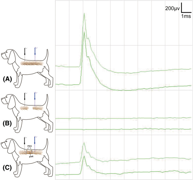
FIGURE 4.

SCEP examination during vASCT. The SCEP examination was conducted in the course of vASCT. Initially, SCEP was performed prior to inducing the spinal cord defect, revealing normal waveforms (A). Subsequently, SCEP was repeated after the creation of the spinal cord defect, resulting in the disappearance of the positive waveform (B). Finally, the conclusive SCEP examination was carried out post‐spinal cord transplantation and topical application of PEG at the interfaces of the two spinal cord surfaces. A positive waveform emerged, albeit with a lower amplitude compared to the SCEP waveform recorded before the induction of the spinal cord defect (C). The stimulating electrode (black) was positioned on the proximal spinal cord surface, while the recording electrode (blue) was placed on the distal spinal cord surface.
