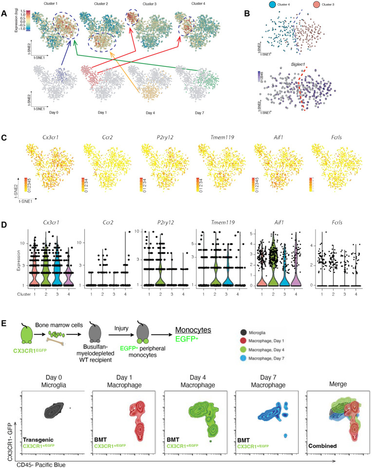
Figure 1. Single-cell RNAseq analysis for retinal CD45+CD11b+ cells 0, 1, 4 and 7 days after ocular injury.
(A) Principal component analysis shows the existence of 4 clusters with distinct transcriptional profiles assigned to the day of cell retrieval. CD45+CD11b+ cells undergo extensive changes in their transcriptome during retinal engraftment and by day 7 acquire an identical signature to naive microglia. (B) Singlec1+ gene is expressed in clusters 3 and 4, both assigned to day 1, suggestive of the presence of monocytes within the microglia sample. (C) tSNE analysis of CX3CR1, CCR2, P2ry12m Tmem119, Aif1, and Fcrls expression in cluster 4 and (D) graphical representation of the expression of microglia markers P2ry12, Tmem119, Aif1, and Fcrls in the 4 clusters, including those representatives of microglia and monocytes signatures. (E) Flow cytometric analysis of the expression of CD45 marker in retinal CX3CR1+ cells before and 1, 4, and 7 days after injury. As reference we used a CX3CR1+/EFGP mouse, stained with CD45 markers. Double positive CD45+ CX3CR1+ cells represent only microglia, since naïve eyes do not have infiltration of monocytes[2]. To map infiltrating/engrafting monocytes, we used a CX3CR1+/EGFP bone marrow chimera. CX3CR1+ CD45+ infiltrating monocytes gradually transitioned their CD45 expression towards the expression of naïve microglia at 7 days.

