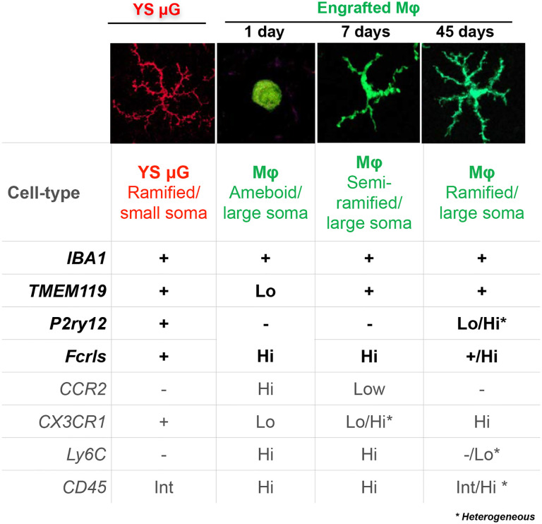
Figure 6. Summary of the microglia and monocyte markers changes.
Monocyte transition from highly amoeboid to highly ramified cells during engraftment into the retina, becoming morphometrically identical and indistinguishable from retinal microglia. These changes are accompanied by suppression of monocyte markers CCR2, Ly6C, and CD45, and upregulation of the tissue-resident macrophage marker CX3CR1+/GFP and microglia markers IBA1, TMEM119, P2ry12, and FCRLS. P2ry12 appears to be conditionally specific to microglia during early infiltration of monocytes) and to a subpopulation of engrafted monocytes.

