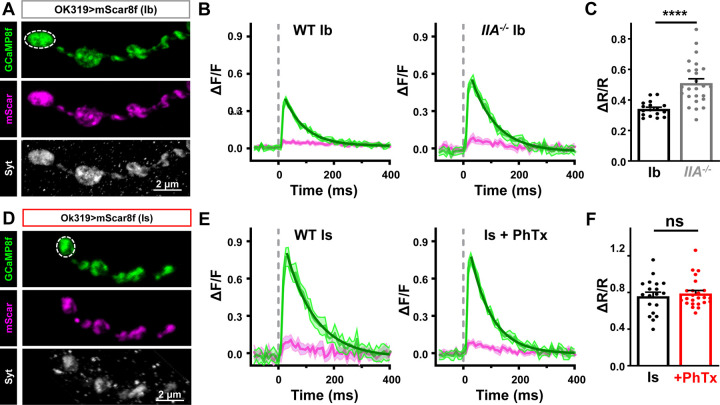
Figure 5: Acute PHP does not enhance presynaptic Ca2+ influx at phasic MN-Is terminals.
(A) Representative immunostaining images of mScarlet and GCaMP8f at MN-Ib (OK319>UAS-Syt::mScarlet::GCamp8f) co-stained with anti-Syt. Note the bouton labeled with dashed lines represents the region of interest undergoing resonant area scanning. (B) Representative Ca2+ imaging traces from MN-Ib resonant area scans of mScar8f expressed in wild type and GluRIIA mutants. Averaged GCaMP8f (green) and mScarlet (magenta) signals from 10 stimuli; shadow indicates +/−SEM. Decays are fit with a one phase exponential (dark green). (C) Quantification of ΔR/R (GCaMP8f/mScarlet Ratio) from MN-Ib boutons demonstrate that chronic PHP enhances Ca2+ signals, as expected. (D-F) Similar images, traces, and quantification as (A-C) but from mScar8f expression at MN-Is in wild type and acute PHP. Note that while baseline Ca2+ levels are higher at MN-Is boutons compared to MN-Ib, as expected, no change is observed after acute PHP signaling. Error bars indicate ± SEM. Additional statistical details are shown in Table S1.

