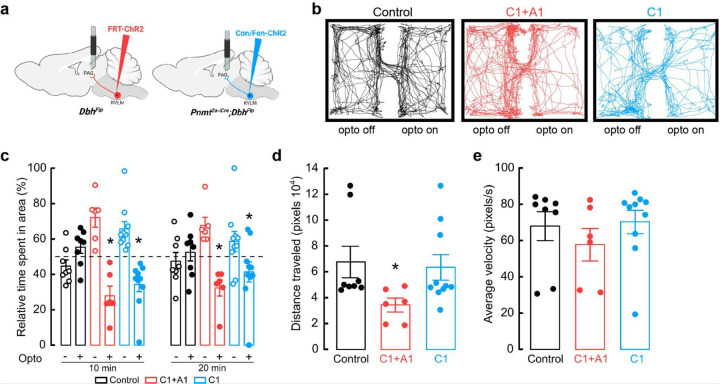
Figure 5. Optogenetic activation of C1 axons innervating vlPAG is aversive.
a, Strategy for targeted viral expression of channelrhodopsin (ChR2) in C1+A1 (red) or C1 (blue) neurons and optic fiber implantation in PAG. b, Representative cumulative trajectories of mice (10 min) in the optogenetic real time place preference (RTPP) assay. Light pulses (20 Hz, 20 ms pulse width) were delivered only when mice inhabited a defined side of the behavioral chamber. c, Average percentage of time spent in the unstimulated (opto ‘off’; open circles) or stimulated (opto ‘on’; closed circles) sides of the chamber. d, Average distance traveled in RTPP assay. e, Average animal velocity during the RTPP assay. Control n=8, C1+A1 n=6, C1 n=10 animals for b-e. Data represents mean ± SEM. *p<0.05 vs control, Kruskal Wallis test with Dunn’s post hoc test.

