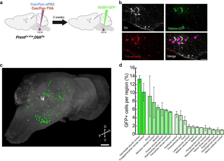
Figure 2. Rabies-mediated transsynaptic tracing identifies presynaptic afferents to C1 neurons.
a, Strategy for intersectional expression of TVA receptor and optimized glycoprotein (oPBG) in C1 neurons to allow rabies-mediated tracing. b, Representative images of C1 starter cells (magenta arrowheads) that are co-labeled by TH (white), GFP (green), and TVA-mCherry (red). Scale bar: 100 μm. c, Representative image of a cleared brain showing manual segmentation of rabies-labeled input neurons (green). d, Average percentage of rabies-labeled neurons detected in specified brain areas. Data represents mean ± SEM. n=3 animals for b-d.

