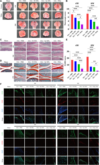
Fig. 9.

Effects of NPs on OA in vivo. Macroscopic observation (A) and corresponding macroscopic scores (B) of OA articular cartilage after 4 and 8 weeks of treatment with the intra-articular injection of NPs. Hematoxylin and eosin staining (C), Safranin O-Fast Green staining (S&F) (D), and corresponding histological scores (E) of articular cartilages after treatment with NPs. (F) The polarization of macrophages in joint synovial membrane was observed by immunofluorescence staining. Macrophages were labeled with F4/80 (green), and M1-type macrophage-related markers were detected by inducible nitric oxide synthase (iNOS) (red), while M2-type macrophage-related markers were detected by CD206 (red). PBS, phosphate-buffered saline; DAPI, 4 DAPI, sphate-buffered salin. Original magnification, ×100. Scale bars, 400 μm. n = 3, *P < 0.05, **P < 0.01, and ***P < 0.001.
