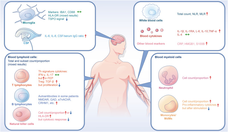
Fig. 1. Immunophenotypes in psychosis.
There are two facets of mismatched systemic changes of immune cells in patients of psychosis. On one facet, blood cytokines and numbers of leukocytes and their innate subsets such as neutrophils and monocytes are increased while activation of innate cells including monocytes and innate lymphoid cells such as NK cells after stimulation are decreased. On the other facet, the mononuclear phagocytic system inside and outside the brain, such as microglia and monocytes, are not synchronically activated in psychosis. That is, there is no overt activation of myeloid cells in the psychotic brain as compared to the immune changes observed in the blood and CSF. α7nAChR α7 nicotinic acetylcholine receptor, CD cluster of differentiation, CRHM1 cholinergic receptor muscarinic 1, CRP C-reactive protein, CSF cerebrospinal fluid, FEP first-episode psychosis, GAD glutamic acid decarboxylase, HLA human leukocyte antigen, HMGB1 high mobility group box 1, IBA1 ionized calcium-binding adapter molecule 1, IFN interferon, IL interleukin, MLR monocyte to lymphocyte ratio, NK natural killer, NLR neutrophil to lymphocyte ratio, NMDAR N-methyl-D-aspartate receptor, S100B S100 calcium-binding protein B, TGF transforming growth factor, TNF tumor necrosis factor, TSPO translocator protein. Green arrows represent no change, blue arrows represent downregulation, and red arrows indicate upregulation.

