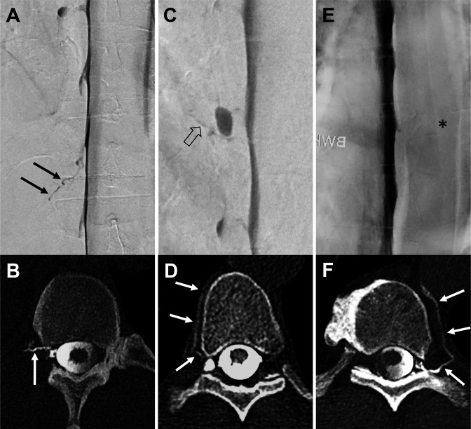
Figure 1.
Three exemplary patients with a combined examination (A+B, C+D, and E+F): lateral decubitus digital subtraction myelography (LD-DSM; A, C, E) in anterior-posterior view followed in each case by lateral decubitus CT myelography (LD-CTM; B, D, F) with a second half of contrast application in axial view. In the first example, a contrasted paraspinal vein at the Th 12/L1 level on the right side is visible in both examinations, LD-DSM (black arrows in A) and LD-CTM (white arrow in B): rated as positive for a CSF-venous fistula (CVF) by raters 1 and 2 in each modality. The second example shows a tiny hyperdense line on LD-DSM (open black arrow in C) adjacent to a nerve root diverticulum at the level of Th 8/9 on the right side (rater 1: CVF-negative, rater 2: CVF-positive, senior rater adjudicated: CVF-positive). On LD-CTM of the same patient, a contrasted paraspinal vein (white arrows in D) is visible at the same level (raters 1 and 2: CVF-positive). The third example does not show a contrasted vein on LD-DSM (asterisk in E) at the level Th 9/10 left (raters 1 and 2: CVF-negative). The LD-CTM of the same patient clearly demonstrates a contrasted paravertebral vein (white arrows in F) at the same level (raters 1 and 2: CVF-positive).

