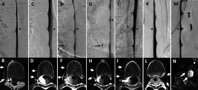
Figure 2.
Seven examples of combined lateral decubitus digital subtraction myelography (LD-DSM) and lateral decubitus CT myelography (LD-CTM) studies (A+B, C+D, E+F, G+H, I+J, K+L, M+N), in which DSM was rated negative but CTM was rated positive for CSF-venous fistulas (CVFs) after senior rating in cases of disagreement between raters 1 and 2. The asterisk in each LD-DSM (A, C, E, G, I, K, M) indicates the spinal level at which a CVF was found at LD-CTM (white arrows in B, D, F, H, J, L, N). Retrospectively, the DSM shows a small lesion as hint of a potential CVF in two examples: a tiny tubular and a dot-like structure in (A) and (I; open black arrows). In example E+F, a patient detected with a CVF only at CTM (F) was treated by transvenous embolization (onyx cast circled by dashed line in G) and was evaluated at follow-up with a de novo CVF at the spinal level above (asterisk in G), also seen only at CTM (white arrows in H). The black arrow in (E) and (G) shows a spinal diverticulum filled with contrast agent only in the lower aspect.

