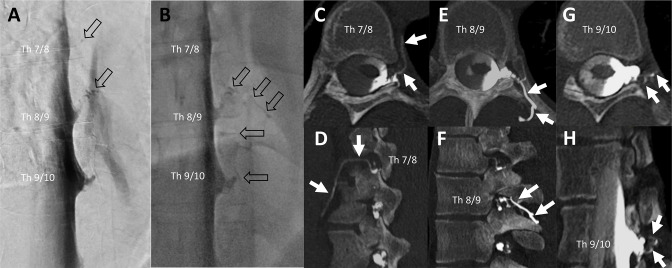
Figure 3.
Example of a patient evaluated as having multiple synchronous CSF-venous fistulas (CVFs; n=3) at the initial examination on the left side (Th 7/8, 8/9, and 9/10). Lateral decubitus digital subtraction myelography (LD-DSM) (A) and lateral decubitus fluoroscopy (B) indicated a CVF at level Th 8/9 by rater 1 and at Th 7/8, 8/9, and 9/10 by rater 2 and the senior rater (open black arrows in A and B). At the level Th 7/8 was a dot-like structure only visible at DSM (open black arrow in A at level Th 7/8). Raters 1 and 2, both identified three CVFs at lateral decubitus CT myelography (LD-CTM) at adjacent spinal levels, shown on axial (C, E, G) and sagittal (D, F) and oblique sagittal (H) CTM by white arrows. As the contrasted veins shown here each have contact with the nerve root sleeve, it is more likely that three CVFs are present than that they originate from one point of fistula only.

