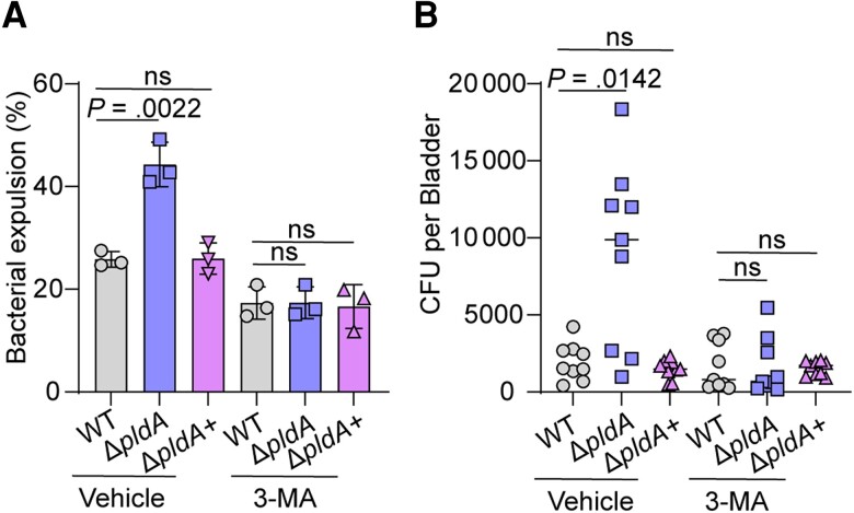
Figure 1.
PldA contributes to UPEC escape from lysosomal exocytosis of BECs. A, Bacterial expulsion of WT, ΔpldA, or ΔpldA+ at 4 hours postinfection in infected 5637 cells pretreated with either vehicle or 2.5 mM 3-MA for 12 hours. B, Bacterial exocytosis into HBSS buffer after 6 hours of incubation with WT, ΔpldA, or ΔpldA+ infected mouse bladders pretreated with or without 3-MA, n = 9. Experiments were repeated 3 times. Data are presented as the mean ± SD. P values were determined using Student t test (A) and Mann-Whitney U test (B). Significance is indicated by the P value. Abbreviations: 3-MA, 3-methyladenine; BEC, bladder epithelial cells; CFU, colony-forming unit; HBSS, Hanks’ balanced salt solution; ns, not significant; UPEC, uropathogenic Escherichia coli; WT, wild type.

