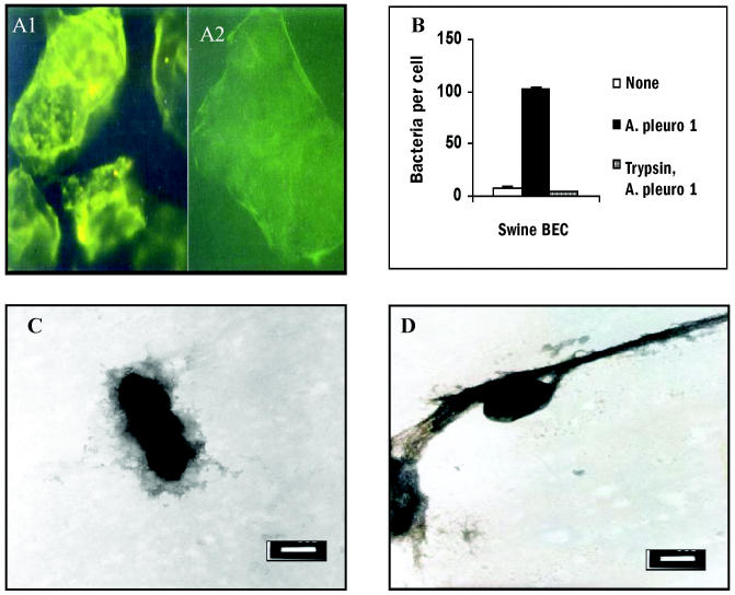
Figure 2.
A1: Swine buccal epithelial cells (BEC) incubated with rabbit anti-swine fibronectin polyclonal antibody and then with anti-rabbit immunoglobulin (Ig)G labelled with fluorescein isothiocyanate (FITC). A2: Effect of trypsin on swine BEC surface; cells were incubated with trypsin (2.5 μg/mL) for 10 min, washed with 0.1 M Tris-HCl, pH 7.2, and treated with the same serum as in A1. B: Numbers of bacteria adhering to swine BEC and to trypsin-treated cells. Assay was performed as in Figure 1. None — indigenous flora; A. pleuro 1 — A. pleuropneumoniae serotype 1. C: Electron micrograph of A. pleuropneumoniae incubated for 5 min with fibronectin and stained with uranyl acetate (bar = 500 nm); bacteria are in close interaction with fibronectin networks, covered by the protein. D: Similar interaction of A. pleuropneumoniae with a fibronectin fibre (bar = 500 nm).

