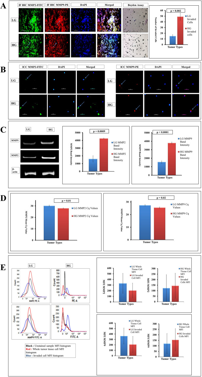
Fig. 2.
a Whole tissue IF-IHC with MMP2-FITC and MMP9-PE contrasted with DAPI (marked with sky blue arrow) suggests increasing expression of both the gelatinases from LG to HG (× 400). The transwell assay also shows a higher number of invaded cells in HG than LG with a significant p value of 0.002 (after 24 h from overlay). b Concurrent higher expression was found in HG tumors in ICC with MMP2-FITC and MMP9-PE along with DAPI (marked with sky blue arrow) (× 400). c Quantitative RNA expression of both the MMP2 and MMP9 shows higher expression in HG than in LG tumors taking β-actin as housekeeping gene with very significant expression profiling (p value is 0.0009 and less than 0.0001, respectively). d Both MMP2 and MMP9 qPCR data shows low Cq values in high grades depicting hyper-expression than low grades (p value 0.01 and 0.02, respectively). e In MFI expression using MMP2-PE and MMP9-FITC, the changes are visible among total and invaded cells. In MMP2, the MFI found in the LG whole tissue cell is higher than invaded sub-type; while in HG, MMP2 expression is higher in invaded sub-type than the whole tissue cell. On the other hand, MMP9 expression also shows similar expression as in LG, the invaded type shows lower expression than whole tissue, while a slight increase in MMP9 expression has been witnessed in invaded cells of HG than its whole cell counterpart (p ≥ 0.05)

