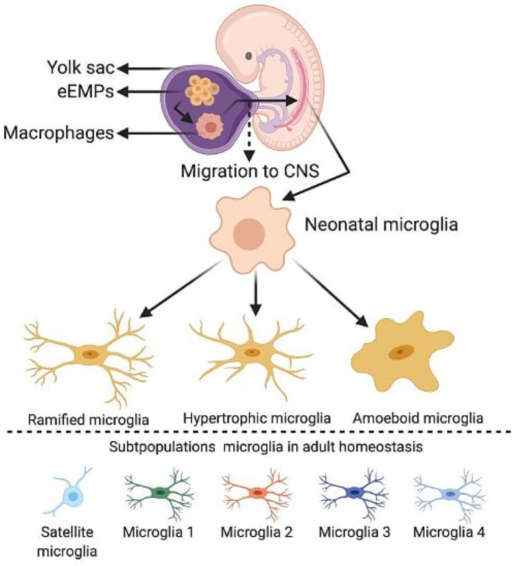
Fig. 1.
Microglia are originated from the early Erythromyeloid Precursors (eEMPs) from the yolk sac embryonic. In the development, they migrate to the neural tube, where they proliferate, colonize the entire parenchyma and remain throughout the life of the organism. Neonatal microglia are characterized by an ameboid morphology with a high rate of proliferation and heterogeneity. In adult brain, microglia are represented by different phenotypes distributed in distinct regions of the CNS that can be identified through different morphologies and molecular markers. The satellite microglia, named due to its location near the neuron, have spherical morphology. These cells interact preferentially in the axon initial segment region. The microglia 1 are identified through the profile of markers: TMEM119+, P2RY12+, CX3CR1+, CD206lo. The microglia 2 are identified through the profile of markers: TMEM119+, P2RY12+, CX3CR1+, CD206lo. The microglia 3 are identified through the profile of markers: expresses TMEM119+, P2RY12+, CX3CR1+, CD11c+, CD68+. The microglia 4 are identified through the profile of markers: TMEM119lo, P2RY12lo, CX3CR1lo, SLC2A5lo, CCL2+, CCL4+, EGR2+, EGR3+. Figure created with BioRender.com

